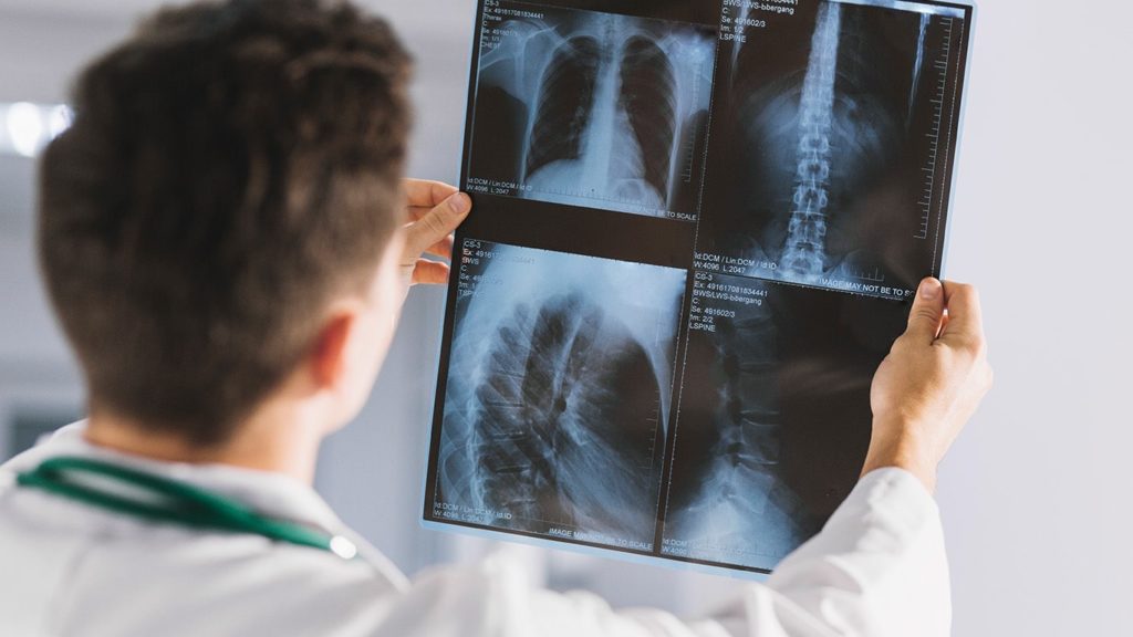
Healthcare professionals need to get into the nitty-gritty of the problems to provide medical assistance. Here is where the X-Rays come into the picture. It helps them to see what’s inside the body and eliminates the need for an incision. Other than this, X-Ray technology is used in the space industry. There, it is proving to be remarkably useful. Currently, industries such as medicine, security, environment, quality control, and manufacturing are using this technology for daily operations.
In layman’s terms, an X-Ray is a type of radiation that is passed through the body, where its energy gets absorbed at different rates. Once the radiation passes, the X-Ray machine turns them into images. And that’s how we can look inside the body for examination.
Even though we get immense benefits from performing X-Rays, the downside is that we are exposed to radiation. The exposure to radiation is low and argued that it does not outweigh the benefits. But we still need to consider several factors before opting for it.
These are the different types of X-Rays:
| X-Ray Type | Primary Usage | Main Focus Area |
|---|---|---|
| Joint X-Ray | Assess joint conditions (arthritis, fractures, infections) | Joints and surrounding bone structure |
| KUB X-Ray | Identify kidney stones or other abnormalities | Kidneys, urinary bladder, ureters |
| Skull X-Ray | Diagnose fractures and diseases, view nasal sinuses | Skull bones and bony structures within the head |
| Dental X-Ray (Intraoral & Extraoral) | Find cavities, check tooth root health, detect impacted teeth, monitor jaw growth | Individual teeth (intraoral), Jaw, and skull (extraoral) |
| Neck X-Ray (Cervical Spine X-Ray) | Diagnose neck pain, injuries, arthritis, disc herniations, tumors | Seven cervical vertebrae and surrounding soft tissues |
| Chest X-Ray | Evaluate lung conditions and heart size | Lungs, heart, ribs, parts of the upper abdomen |
| Pelvis X-Ray | Identify fractures, infections, tumors, hip dysplasia, arthritis | Lower body structure, hips, upper thighs |
1. Joint X-Ray
In cases of bone fracture, and wear and tear of cartilage or ligament, the Joint X-Ray is brought up. The reason is, that it shows enough details to highlight the problem. In this type, the X-Ray is taken of the shoulder, wrist, hip, knee, and ankle.
The purpose of performing this test is to identify the cause of difficulties in your joints. It is usually used to detect arthritis, fractures, degenerative bone conditions, or tumors in the joints. The other name for this test is Arthrography and Arthrogram.
Even though the exposure to radiation is low and outweighs the benefits, it can still put you at risk. And that’s why you will be guided to wear a protective shield to cover the areas that are not being scanned.
Joint X-Ray Overview:
- Used to assess joint conditions like arthritis, fractures, or infections.
- Can detail the bone surfaces near the joint and the space between the bones.
- Doesn’t show muscles, ligaments, or cartilage well.
2. Kidney, Ureter, and Bladder X-Ray
When patients come with severe problems of abdominal pain, then the Kidney Ureter and Bladder X-ray are suggested. It helps to diagnose the cause of problems. Along with this, it is used to diagnose urinary tract disorders, gallstones, and kidney stones.
Firstly, the x-ray assesses the urinary tract, obtaining the size, shape, and positions of the organs. These organs are the kidneys, ureters, and bladder. Then, the X-Ray images of the digestive system are taken. Overall, this test helps medical professionals to diagnose conditions such as tumors, intestinal blockage, and foreign objects in the stomach.
Kidney, Ureter, and Bladder X-Ray (KUB) Overview:
- Utilized to identify kidney stones, although it can also detect some other abnormalities.
- It shows the size, shape, and position of the kidneys, bladder, and the path of the ureters.
- It does not use contrast dye and cannot provide detailed images of internal kidney structures.
3. Skull X-Ray
There are several reasons for which the Skull X-Ray is performed. A few of these reasons are diagnosing tumors, head injuries, fractures, deformities, and decalcification. Here, along with the bones of the skull, the facial bones are examined.
The X-Ray images are taken from different views, including front and side views. The duration of the test is not more than 20 to 30 minutes. Even though we have advanced technology such as Magnetic Resonance Imaging(MRI) and Computed Tomography(CT), the Skull X-Ray is used for detecting conditions of the skull and brain.
Skull X-Ray Overview:
- Makes visible the bony structure of the head.
- Often used to diagnose fractures and disease, to view the nasal sinuses, or to evaluate cranial bones.
- Does not provide a detailed view of the brain.
4. Dental X-Ray
In dental X-Ray, the images are captured for identifying teeth, gum, and jaw problems. To be specific, it is used to identify cavities, tooth decay, jaw bone loss, abscesses, and cysts. There is bone loss in cases of gum disease, which is also revealed in this type of X-Ray. Depending on the X-Ray film position, it is classified into two types: Intraoral and Extraoral.
The Intraoral includes Bitewing, Occlusal, and Periapical. Bitewing X-Ray is used for teeth above the gum line, and it detects the thickness of the bone caused by diseases. Occlusal X-Ray is used for the roof or floor of the mouth and helps to track the placement and development of the teeth in the jaw. Periapical X-Ray shows the complete teeth from the root to the crown, and it is used to detect unusual changes in the root.
The Extraoral includes Panoramic, Cephalometric, and Sialogram. Panoramic X-Ray shows the entire mouth and is used to diagnose tumors. Cephalometric X-Ray shows the side of the face, and it is recommended for different conditions and injuries. Additionally, it is used to develop treatment plans. The Sialogram X-Ray shows salivary ducts and glands, and it is used to detect salivary gland problems.
Dental X-Ray Overview:
Intraoral X-Ray:
- Provides detailed images of individual teeth.
- It also helps with finding cavities, checking the health of the root and bone surrounding the tooth, and monitoring tooth development.
Extraoral X-Ray:
- Primarily focuses on the jaw and skull.
- Less detailed than intraoral x-rays, but helpful in detecting impacted teeth, and monitoring growth and development of jaws. This type of x-rays also helps in identifying problems between teeth and jaws and the temporomandibular joint (TMJ).
5. Neck X-Ray
The neck X-Ray captures the images of the cerebral vertebrae, which includes the seven bones of your spine in the neck. The other name of this x-ray is cervical spine x-ray. It is used to detect neck injuries, osteoporosis, dislocation, inflammation, foreign objects, and disk problems. Since the neck supports the weight of our head, it is particularly vulnerable in cases of accidents and falls. So in these situations, a neck x-ray is performed.
Neck X-Ray (Cervical Spine X-Ray) Overview:
- They aim to visualize the seven cervical vertebrae and surrounding soft tissue.
- Useful in diagnosing neck pain, injury, arthritis, disc herniations, tumors, and infections.
- Cannot see nerves or spinal cord clearly.
6. Chest X-Ray
When patients come with problems of chest injury, chest pain, or shortness of breath, they are suggested to get a Chest X-Ray. Here, the images of the heart, lungs, chest bone, and spine are produced. It helps to diagnose serious conditions such as pneumonia, lung cancer, heart problems, broken ribs, and emphysema. It also reveals the size and outline of the heart, which indicates heart failure.
Additionally, it detects calcium deposits and blood vessel problems. Certain medical conditions change the structure of the heart or lungs. So it becomes essential to perform a chest x-ray.
Chest X-Ray Overview:
- Primarily used to evaluate lungs but also provides information about the heart, ribs, and parts of the upper abdomen.
- Can spot pneumonia, heart failure, lung cancer, tuberculosis, and other lung conditions.
- Will not provide detailed images of the inside of the heart or the bronchi.
7. Pelvis X-Ray
The pelvis bone image is captured in this type of X-Ray including hip bones, sacrum, and coccyx. It is used to detect conditions such as pelvic fractures, tumors, hip dislocations, and arthritis. A Pelvis X-Ray is used to plan in cases where surgery is required. Apart from these reasons, a Pelvic X-Ray is required to detect cysts and infections. This test is performed in times of traumatic events, which may include accidents and falls.
Since the injured patients are exposed to radiation, they may experience some pain during the process. But overall, as mentioned, the benefits of X-rays outweigh the risks. It helps us to identify the cause of the problem and start the treatment as soon as possible.
Pelvis X-Ray Overview:
- Take the image of the lower body structure including – the hips and the upper portion of the thighs.
- It can identify fractures, infections, tumors, hip dysplasia, arthritis, and other abnormalities.
- Does not detail soft tissues like muscles or internal organs well.